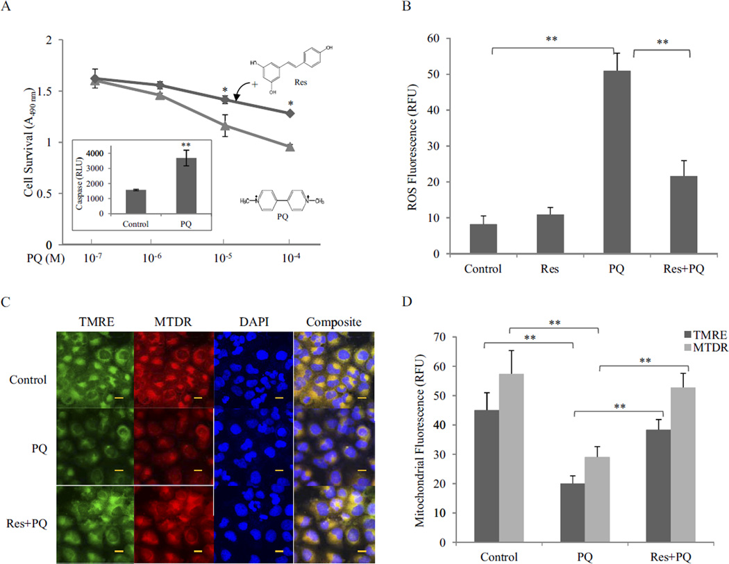
Fig. 1.
Inhibition of PQ toxicity by Res. A, cytotoxicity. BEAS-2B cells were treated with PQ or PQ + Res (10 µM) for 24 h in triplicate. Cytotoxicity was detected by using CellTiter 96 AQueuos One Solution Reagent. Data represent means from three samples. Inset, apoptosis (caspase 3/7 activation) was measured in cells treated with PQ at 10 µM for 24 h. Caspase activity was expressed in RLU. B, ROS production. Cells were treated with Res (10 µM) for 16 h then with or without PQ (20 µM) for 24 h. ROS production was measured by using fluorophore DHE and expressed in RFU. Data represent means ± S.D. from three samples. C, mitochondrial damage. Cells were treated as in B. Mitochondrial damage was assayed by using fluorophores TMRE (green) and MTDR (red). DAPI (blue) was used to counterstain the nucleus. Composite images show the mitochondria in yellow (merge of green and red). Bar size, 20 nm. D, quantification of TMRE and MTDR fluorescence.*, p < 0.05; **, p < 0.01.

