Fig. 3.
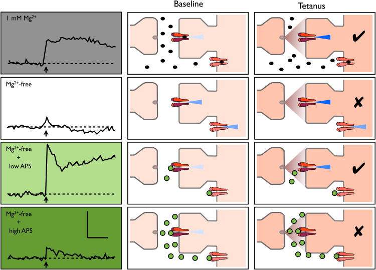
Inappropriate activation of NMDARs inhibits LTP. The panels show data (left panel; replotted from Coan et al, 1989) and schematics during baseline (centre) and a tetanus (right) under four experimental conditions (from top to bottom): in 1 mM Mg2+ (grey shading and black circles), following perfusion with Mg2+-free medium, and following the addition of either 20 μM or 200 μM D-AP5 in Mg2+-free medium (green shading and circles). Calibration bar is 4 mV and 5 min. Optimal conditions for LTP requires minimal activation of NMDARs except during the induction stimulus (time of delivery of the tetanus is indicated by an arrow). By removing Mg2+, NMDAR activation in enhanced throughout the recording period and this inhibits the generation of LTP. A low concentration of D-AP5 normalizes the situation by inhibiting spurious NMDAR activation, but is outcompeted by L-glutamate during high frequency stimulation. However, a high concentration of D-AP5 inhibits NMDARs during high frequency stimulation.

