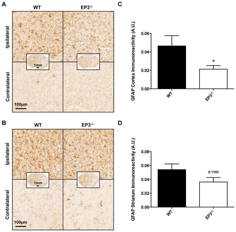
Fig. 5.
Effect of PGE2 EP3 receptor deletion on astrogliosis after ICH. Immunohistochemical staining for GFAP was used to evaluate cortical and striatal astrogliosis in WT and EP3−/− mice 72 h following ICH. (A and B) Representative high magnification images of coronal brain sections showing the ipsilateral and contralateral (A) cortex and (B) striatum for WT (left) and EP3−/− mice (right). Square selections in the inserts denote magnified regions. (C and D) Quantification of brown positive pixel count demonstrated that EP3−/− mice had significantly reduced (C) cortical GFAP immunoreactivity, and tended to have less (D) striatal astrogliosis. Both groups demonstrated negligible staining in the contralateral cortex and striatum; thus, data is presented as ipsilateral immunoreactivity corrected for the area of quantification, without normalization for the contralateral equivalent (see methods and supporting information). All comparisons included n = 7 WT and n = 8 EP3−/− mice and *p < 0.05.

