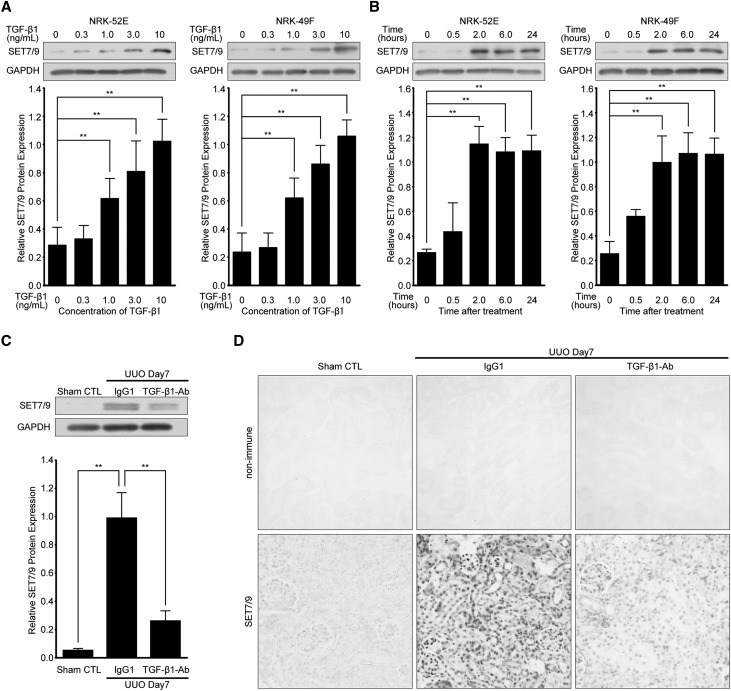
Figure 2.
TGF-β1 induces SET7/9 expression in renal cells, and injection of neutralizing TGF-β1 antibody reduces SET7/9 expression after UUO. NRK-52E cells and NRK-49F cells were treated with TGF-β1. Representative western blot analysis showing the levels of SET7/9 protein expression in TGF-β1-stimulated NRK-52E cells and NRK-49F cells at (A) various dosages (time; 24 hours) and (B) time points (TGF-β1; 10 ng/mL). Expression levels were compared with vehicle-treated control. Quantification is shown in the lower panel. Data were analyzed by one-way ANOVA followed by Dunnett post hoc test based on vehicle-treated controls (n=5 for each group). (C) Representative western blot analysis with anti-SET7/9 antibody. Quantification is shown in the lower panel. Data were analyzed by one-way ANOVA followed by Dunnett post hoc test based on UUO mice with control IgG1 injection (n=5 for each group). (D) Images of SET7/9 staining demonstrating the levels of SET7/9 expression by intraperitoneal injection of neutralizing TGF-β1 antibody (TGF-β1-Ab) at a dose of 1.5 mg/kg/48 hours compared with control IgG1 at the same dose of TGF-β1-Ab. Original magnification, ×200. **P<0.01. Sham CTL, sham-operated controls; IgG1, UUO mice with control IgG1 injection; non-immune, control non-immune serum; TGF-β1-Ab, UUO mice with neutralizing TGF-β1 antibody injection.

