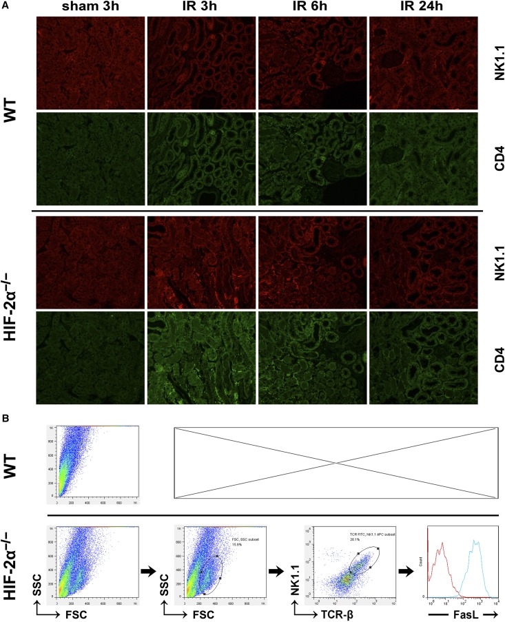
Figure 3.
HIF-2α KO promoted infiltration of NKT cells into ischemic kidneys. (A) HIF-2α−/− and WT mice were subjected to 20 minutes of renal ischemia. Renal sections from sham-operated mice or ischemic kidneys harvested at 3, 6, and 24 hours after ischemic insult were stained with the indicated antibodies, followed by confocal microscopic analyses. Representative photographs are shown. Original magnification, ×200. Similar results were obtained in six independent experiments and summarized in Table 2. (B) HIF-2α−/− mice and WT littermates were subjected to 20 minutes of bilateral renal ischemia. Both kidneys were harvested at 3 hours after reperfusion and the infiltrating inflammatory cells were isolated by using CD45 microbeads, stained with antibodies against NK1.1 (APC), TCR-β (FITC) and FasL (PE), and subjected to FACS analysis, as described in the Concise Methods. The expression of FasL was analyzed on electronically gated NK1.1+TCR-β+ (NKT) cells. The blue line indicates the staining of FasL antibody, and the red line indicates the background staining with isotype-matched control IgG. Similar results were obtained in six independent experiments and a representative photograph are shown.

