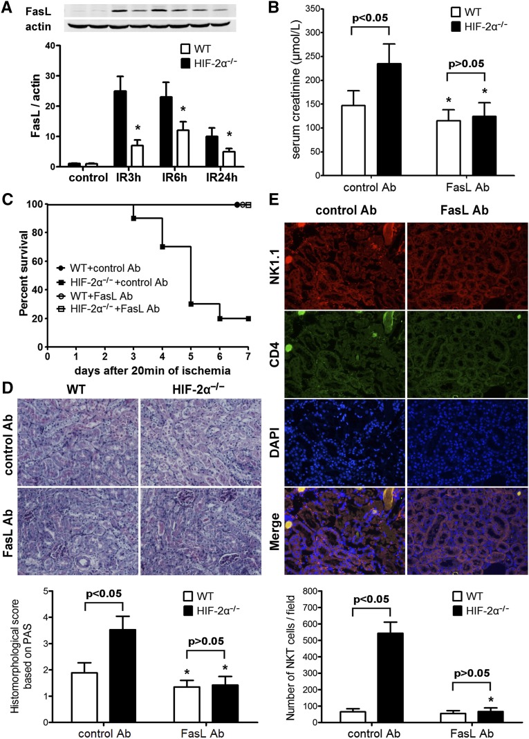
Figure 5.
Expression of FasL in post-ischemic kidneys and the effect of FasL blockade on renal IR injury. (A) HIF-2α−/− and WT mice were subjected to 20 minutes of renal ischemia. Renal samples were harvested at 3, 6, and 24 hours after reperfusion. The kidneys from sham-operated mice served as controls. Expression of FasL was evaluated by western blot analysis and co-detection of β-actin was performed to assess equal loading (n=4 for each group at each time point). FasL protein bands were quantified and normalized to β-actin. *P<0.05 versus HIF-2α−/− samples. (B) The mice were subjected to FasL Ab or control Ab pretreatment and 20 min of renal ischemia. Serum creatinine levels at 24 h after the reperfusion were shown (n=6 per group). *P<0.05 versus isogenic mice treated with control Ab. (C) Survival of mice after FasL Ab/control Ab pretreatment and 20 min of renal ischemia (n=10 per group). Compared with control Ab, FasL Ab led to a significant survival advantage in HIF-2α−/− mice by Kaplan-Meier analysis (log-rank test, P<0.05). (D) Representative PAS-stained sections in post-ischemic kidneys harvested at 24 h. Original magnification, ×200. Abnormalities based on PAS-stained sections were graded and data are expressed as mean±SD from 6 mice per group. *P<0.05 versus control Ab-treated isogenic mice. (E) Renal sections from ischemic kidneys harvested at 3 hours after reperfusion were stained with the NK1.1 and CD4 antibodies, followed by confocal microscopic analyses. Representative photographs from HIF-2α−/− mice are shown. Original magnification, ×200. A summary of the quantitative analysis of double-positive infiltrating cells per field was presented. Data are expressed as mean±SD from 4 animals per group. *P<0.05 versus control Ab-treated HIF-2α−/− mice. PAS, periodic acid–Schiff.

