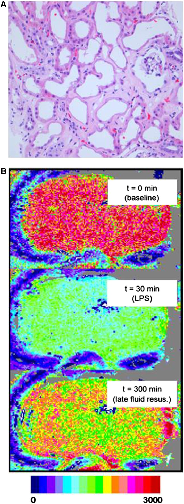
Figure 2.
(A) Histopathology of human kidney biopsy from the cortical area in a patient with AKI. Note the enlarged interstitial space with markedly reduced peritubular capillaries, ongoing cellular plugging of existing capillaries, deteriorating tubules, and an ischemic shrunken glomerulus. This is a later stage of ischemic AKI likely resulting in microvascular dropout, CKD development, and/or acceleration of CKD progression. Reprinted from Steve Bonsid (Nephropath, Little Rock, AK), with permission. (B) Shown is a speckle imaging perfusion map of the surface of a rat kidney61 at baseline, during septic shock, and after fluid resuscitation after a 30-minute delay. Fluid resuscitation corrected systemic hemodynamic variables but induced heterogeneous areas of hypoperfusion in the renal cortex. Reprinted from Legrand et al.,40 with permission.

