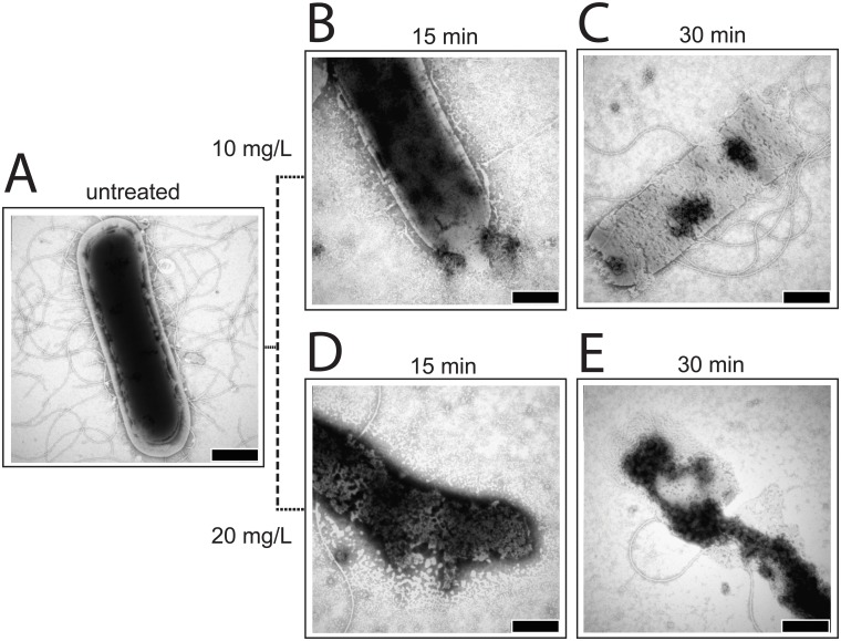
Fig 3. TEM pictures of B. subtilis cells.
0.25% phosphotungstic acid at pH 7.3 was used for staining. A. Untreated. B. Treated with 10 mg/L of DR5026 for 15 min. C. Treated with 10 mg/L of DR5026 for 30 min. D. Treated with 20 mg/L of DR5026 for 15 min. E. Treated with 20 mg/L of DR5026 for 30 min. The scale bars in the right-hand corners of the pictures represent 500 nm.

