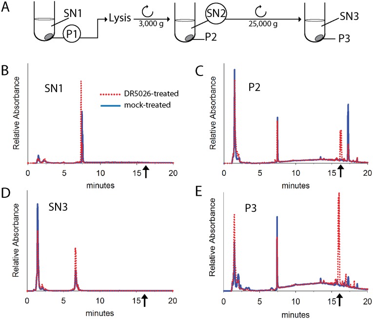
Fig 4. Localization of DR5026 in B. subtilis cells.
A. A scheme of the experiment. SN, supernatant; P, pellet. B. HPLC data of supernatant after cell sedimentation, SN1. C. HPLC analysis of cell debris and remaining non-lysed cells, P2. D. HPLC analysis of cell cytoplasm, SN3. E. HPLC analysis of the plasma membrane fraction, P3. The dotted red line: DR5026 treated cells; the blue line: mock-treated cells. The arrows indicate where DR5026 eluted from the column. The identity of DR5026 was confirmed by MS detection.

