
Figure.
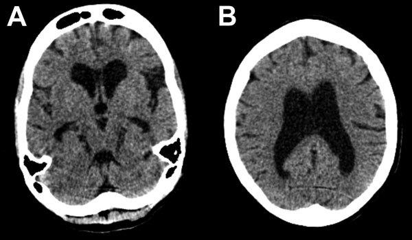
Representative axial cuts from noncontrast head computed tomography scan imaging of a 30-year-old woman with encephalitis resulting from Ebola virus infection, Sierra Leone. Images show global atrophy in keeping with nonobstructive ventriculomegaly and no periventricular low attenuation: A) subcortical atrophy; B) cortical atrophy. There was no evidence of hydrocephalus, previous stroke, or intracranial hemorrhage. A cavum septum pellucidum was noted in other images.