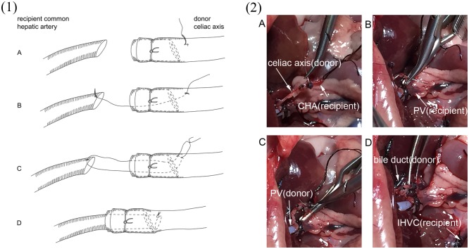
Fig 3. Depiction of the procedure of vessel anastomosis between donor celiac axis and recipient common hepatic(CHA).
The vessel end of the recipient CHA and the donor celiac axis were placed closely to facilitate the anastomosis. (A): The first suture was performed with a length of 10–0 silk from outside to inside of the donor celiac axis and the suture was placed approximately 1 mm beyond the bracket. (B): The suture went transmurally through the anterior edge of the recipient CHA. (C): The donor celiac axis was repierced from inside to outside near the first suture point by the same thread. (D): Finally, the thread was pulled gently to guide the recipient CHA onto the donor celiac axis, and the suture was tied. Fig 3-(1) is diagram illustration showing the 4 steps of anastomosis. Fig 3-(2) shows the 4 steps with real photos of surgical procedure.

