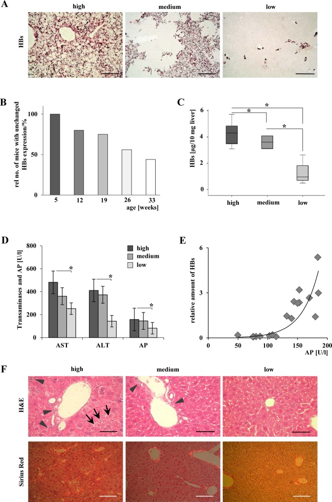
Fig 1. Loss of HBs expression ameliorated liver injury.
(A) Immunohistochemical staining of HBs demonstrated the appearance of hepatic areas without transgene expression in distinct mice beginning with the age of 12 weeks. At the same age, mice with HBs expression in single hepatocytes and mice with expression of HBs in every hepatocyte were observed. Representative micrographs are shown. Scale bars in left and right panels: 160μm, magnification x200. Scale bar 320 μm in the medium panel: magnification x100. (B) The observed loss of HBs expression is age-dependent. The relative number of mice exhibiting areas without HBs expression increased with higher age of the mice. Bars indicate the relative number (%) of animals with unchanged HBs expression in all hepatocytes. (C) Quantitative analysis of HBs expression in liver tissue assessed by ELISA. *P < 0.05. (D) Amelioration of liver serum parameters correlates significantly with loss of HBs expression. *P < 0.05. (E) Correlation of AP and HBs. r2 = 0.864, P < 0.001. (F) H&E staining indicates ground glass hepatocytes and enhanced numbers of inflammatory cells in the „high”and „medium”group. Scale bars 80 μm, magnification x400. Arrowheads: inflammatory infiltrates, arrows: ground-glass-hepatocytes. Sirius Red staining indicates fibrillary collagen. Scale bars 160μm, magnification x200.

