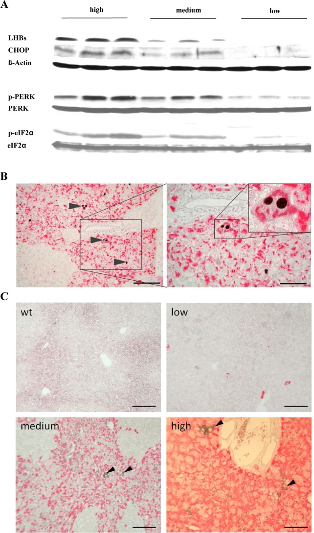
Fig 2. Spontaneous loss of HBs expression reduced ER-stress and apoptosis.
A) Representative Western blots of p-eIF2α, p-PERK, and CHOP demonstrated the correlation between HBs expression and activation of ER-stress. Loading controls: ß-Actin, eIF2α, and PERK. (B) Co-immunostaining of HBs (red) and CHOP (black) in transgenic mouse liver. The micrographs demonstrate that HBs expressing hepatocytes undergo apoptosis only. Scale bars 160/80 μm. Arrowheads indicate CHOP stained apoptotic hepatocytes. (C) Co-immunostaining of HBs (red) and GRP78 (black) in transgenic mouse liver. IHC demonstrated strong expression of GRP78 in selected hepatocytes in centrilobular areas. Scale bars 50 μm. Arrowheads indicate GRP78 stained hepatocytes.

