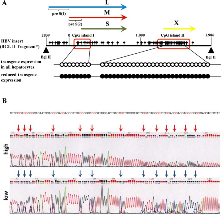
Fig 4. Methylation analysis of HBV-genomic sequence.
A) Mice with reduced amount of transgene expression exhibited methylated CpG islands I+II (red boxes, figure modified from [12]). Unfilled circles indicate non-methylated CpG sites. Presence of methylated CpGs is indicated by black filled circles and was detected by analyzing six independent mice with reduced transgene expression. L, M, and S indicate the sequences encoding the large, middle, and small surface proteins. X indicates the sequences encoding the X-protein. *HBV insert (Bgl II fragment) according to Chisari et al. 1986 [30]. B) A representative result of Bisulfite sequenced CpG island II demonstrates methylation of all CpG sites in mice with reduced HBs expression (low).

