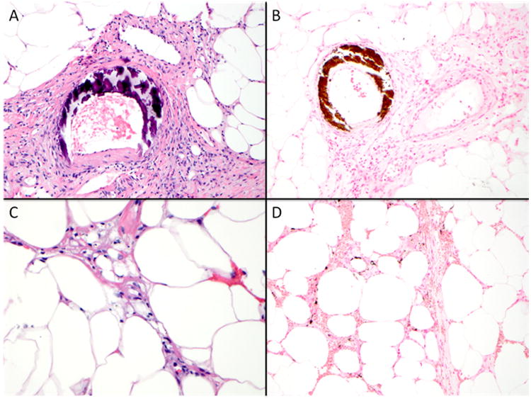
Figure 2. Histopathology of calciphylaxis.

Course basophilic medial calcification of small arteries as demonstrated by Hematoxylin & Eosin stain (400×) and highlighted by von Kossa histochemical stain (200×) (Panel A-B). Septal panniculitis and subcutaneous fat necrosis with presence of subtle finely granular basophilic calcium deposits (400×, Hematoxylin & Eosin, Panel C). A von Kossa histochemical stain aids in the detection of interstitial calcium deposits, which may not be identified on routine histologic sections (200×, Panel D).
