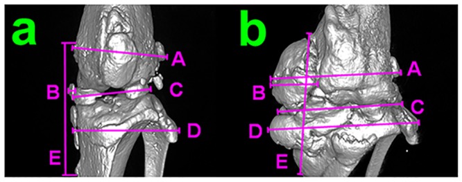
Fig 2. Joint grading.

a) cranial view of a 3D reconstruction of uCT data from a joint with minimal perturbations. b) cranial view of a 3D reconstruction of uCT data from a joint with maximal perturbations. Measured parameters include A-distal femur width, B-medial collateral ligament thickness, C-joint width at the articulating surface, D-proximal tibia/fibula width, E-medial collateral ligament length.
