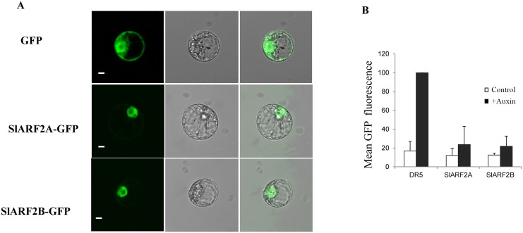
Fig 3. Subcellular localization and functional analysis of SlARF2A and SlARF2B by single cell system.
(A) Subcellular localization of tomato SlARF2A/2B proteins. SlARF2A/2B-GFP fusion proteins were transiently expressed in BY-2 tobacco protoplasts and subcellular localization was analyzed by confocal laser scanning microscopy. The merged pictures of the green fluorescence channel (left panels) and the corresponding bright field (middle panels) are shown in the right panels. The scale bar indicates 10 μm. The top pictures correspond to control cells expressing GFP alone. The middle and bottom pictures correspond to cells expressing the SlARF2A-GFP and SlARF2B-GFP fusion proteins, respectively. (B) SlARF2A/2B protein represses the activity of DR5 in vivo. SlARF2A/2B proteins were challenged with a synthetic auxin-responsive promoter called DR5 fused to the GFP reporter gene. A transient expression assay using a single cell system was performed to measure the reporter gene activity. Tobacco protoplasts were transformed either with the reporter construct (DR5::GFP) alone or with both the reporter and effector constructs (35S::SlARF2A/2B) and incubated in the presence or absence of 50 μM 2,4-D. GFP fluorescence was measured 16 h after transfection. For each assay, three biological replicates were performed. GFP mean fluorescence is indicated in arbitrary unit (a.u.) ± standard error.

