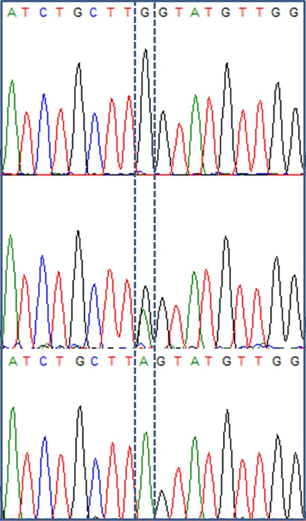
Fig 3. Electropherogram showing G/A mutation at nucleotide 1084 (located on the last nucleotide of exon 3).
The nucleotide of interest is marked by a dotted box. The upper panel shows data from a subject homozygous for wild-type G nucleotide, the middle panel shows a subject with unconjugated hyperbilirubinemia who had heterozygous GA genotype, and the lower panel shows a subject with unconjugated hyperbilirubinemia who had homozygous mutant A genotype.

