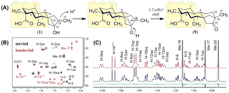
Figure 5.
(A) Wagner–Meerwein rearrangement of steviol (1) to form isosteviol (9), highlighting the conserved fragment C-1/C-7. (B) Comparison between sections of the 2D 1H,13C-HSQC experiments of 1 (in black) and 9 (in red). (C) Section of the calculated (in red) and experimental (in blue) 1H NMR spectra of 9 in DMSO-d6 (37 mM, 600 MHz, 298 K). Calculated residuals are shown in green.

