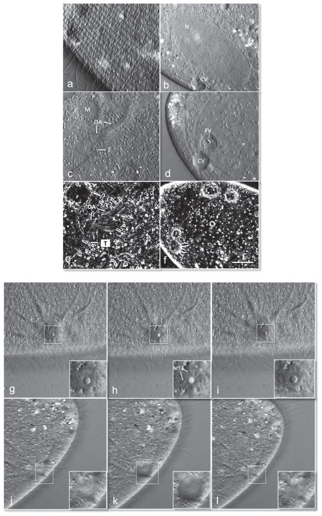
Fig. 1.
High resolution images of an immobilized paramecium. Differential interference contrast (DIC) images of paramecium using an Olympus 100X objective (a–d). (a) Paramecium cortex highlighting the ciliary row with basal bodies and kinetodesmal fibers. (b) Parental macronucleus (N) and contractile vacuole (CV) in diastole. (c) Oral apparatus undergoing morphogenesis. The trichocysts (T) and mitochondria (M) are labeled. Also see Video 1. (d) Filled CV prior to discharge and food vacuole (FV) filled with bacteria. Orientation-independent DIC (OI-DIC) images of paramecium using 100X objective (e and f). (e) OI-DIC image of same cells shown in (c) a few seconds later. (f) Filled CV prior to discharge of contents. Image series of cycle of CV opening and closing top down view, inset corresponds to dotted box within image (g–i). (g) Closed pore within CV, (h) opening of the pore within the CV and (i) closing of the pore to complete one cycle of systole. The microtubules (MT) extend from the pore and radiate towards the canals. Image series of side view of CV and pore through cycle of diastole (j–l). The pore gradually fills its contents (j and k) and then releases them (l). Also see Videos 2 and 3. Scale bar is 10 μM.

