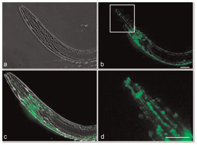
Fig. 4.
Phase contrast and epifluorescent images of neural network within immobilized Caenorhabditis elegans. The nematode worm was imaged using phase contrast (a) and epifluorescent imaging (b). The neural network within the head and back fluoresce. The overlay of the phase and fluorescent images is shown in (c). (d) Close up view of neural network in head as outlined in white box in (b). Also see Video 10. Scale bar is 10 μM.

