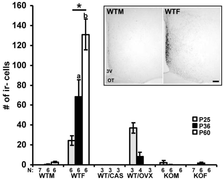
Fig. 1.

A digital inset image shows representative sections with kisspeptin-ir cells in the AVPV at P60 in WTM and WTF mice. The graph illustrates the number of kisspeptin-ir cells in the AVPV in gonadally intact wild type (WT) male (WTM) and female (WTF), gonadectomized at P21 WT male (WT/CAS), and female (WT/OVX), and agonadal SF-1 KO male (KOM) and female (KOF) mice in three different developmental stages, at P25 (before puberty), at P36 (during pubertal time) and at P60 (adult). The number of cells is presented as mean +/- SEM. *p < 0.05 (significantly different from the WTM, WT/CAS, WT/OVX, KOM and KOF); ap< 0.05 (significantly different from WTF at P25); bp< 0.05 (significantly different from WTF at P36). For the image, 3V- third ventricle, OT- optic tract, bar: 100 μm.
