
Fig. 1.
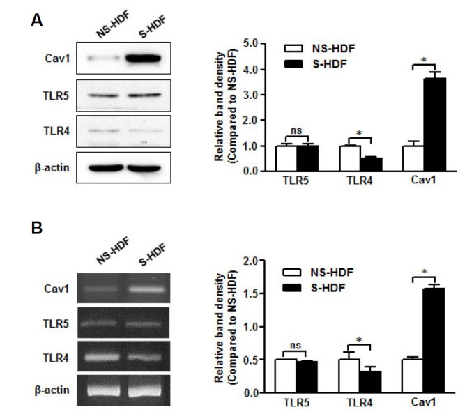
TLR5 is conserved in senescent non-immune cells. RNA and protein were isolated from NS-HDF and S-HDF cells. (A) Caveolin-1, TLR4, and TLR5 protein expression was detected by Western blot with specific antibodies. (B) Caveolin-1, TLR4, and TLR5 mRNA levels were analyzed by RTPCR with specific primers. The β-actin was used as the loading control for both RT-PCR and Western blotting. The relative density of protein expression and mRNA levels were normalized to β-actin and represented by quantitative graphs. Data are presented as mean ± SD from five independent experiments; *p < 0.05; ns, not significant.