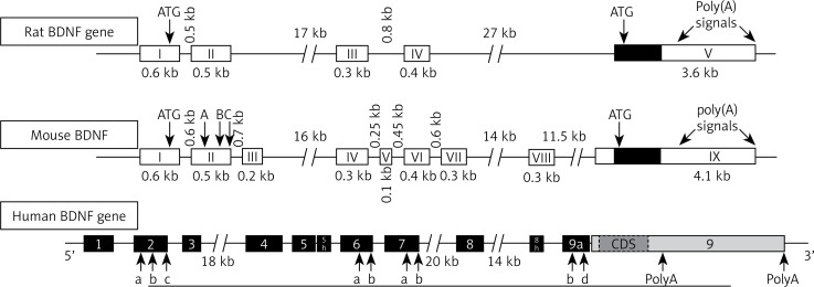
Figure 2.
Gene structure of BDNF. Note the presence of four promoters in rat and 9 promoters in mouse. Each of the driving transcripts of BDNF mRNAs containing one of the four 5′ non-coding exons (I, II, III, IV) in promoters is later spliced to the common 3′ protein coding exon. Human BDNF structure and its splicing variant are seen above with arrows indicating alternative polyadenylation sites (PolyA) in the 3′-UTR and internal alternative splice sites in exons 2, 6, 7 and 9a (letters a, b, c and d) [18]. Arrangement of introns and exons on BDNF genes is determined by analyzing genomic and mRNA sequence using bioinformatics, RACE, and RT-PCR [17]

