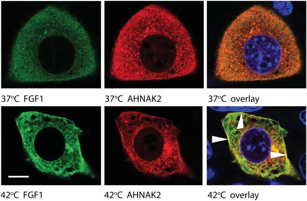
Figure 3. Heat shock induces a peripheral localization of FGF1 and ctAHNAK2.
NIH 3T3 were adenovirally transduced with FGF1:HA and ctAHNAK2:V5 and plated on glass coverslips. Adherent co-transduced cells were incubated in serum-free medium at 37°C or stressed at 42°C for 90 min and then formalin fixed. Cells were co-stained with FITC-labeled anti-HA antibodies (green), CY3-labeled anti-V5 antibodies (red), DAPI (blue), and studied via a confocal microscope. Scale bar indicates 8 μm. Arrows indicate AHNAK2 and FGF1 colocalization at the periphery of a heat shocked cell.

