
Figure 10.
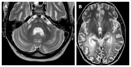
Adrenoleukodystrophy. A: Axial T2 image shows symmetric increased T2 signal in the MCP and bilateral corticospinal tracts; B: Axial T2 show concomitant symmetric and confluent white matter abnormal signal in bilateral occipito-parietal regions, typical for ADL. Note the preservation of subcortical u-fibers. Images courtesy of Dr. Lily Wang. ADL: Adrenoleukodystrophy; MCP: Middle cerebellar peduncles.