
Figure 12.
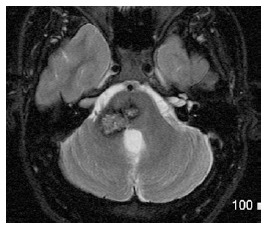
Cavernoma of middle cerebellar peduncle. Axial T2-WI shows contiguous lesions involving the pons and right MCP, with a typical mixed speckled hyper-intense and hypo-intense center and peripheral halo of hypo-intensity due to chronic hemosiderin deposition. MCP: Middle cerebellar peduncles.