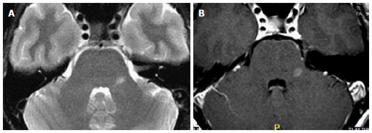
Figure 2.

Multiple sclerosis. A 40-year-old female with 2 mo history of left facial pain/numbness and difficulty talking (scanning speech). Axial T2 (A) and axial post-contrast MR (B) images show isolated lesion in the left MCP. Due to suspicion for demyelinating disease MRI of the spine and CSF analysis was recommended. Additional lesion found in the thoracic cord (not shown) and CSF led to the diagnosis of MS. MCP: Middle cerebellar peduncles; CSF: Cerebrospinal fluid; MRI: Magnetic resonance imaging; MS: Multiple sclerosis.
