
Figure 7.
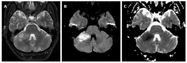
Anterior inferior cerebellar artery infarct. Patient with acute cerebellar ataxia and right-sided weakness. Axial T2 (A), DWI (B) and ADC maps (C) show well-defined area of high T2 signal and restricted diffusion consistent with ischemia. DWI: Diffusion weighted-imaging.