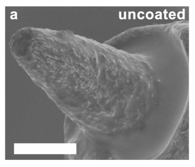
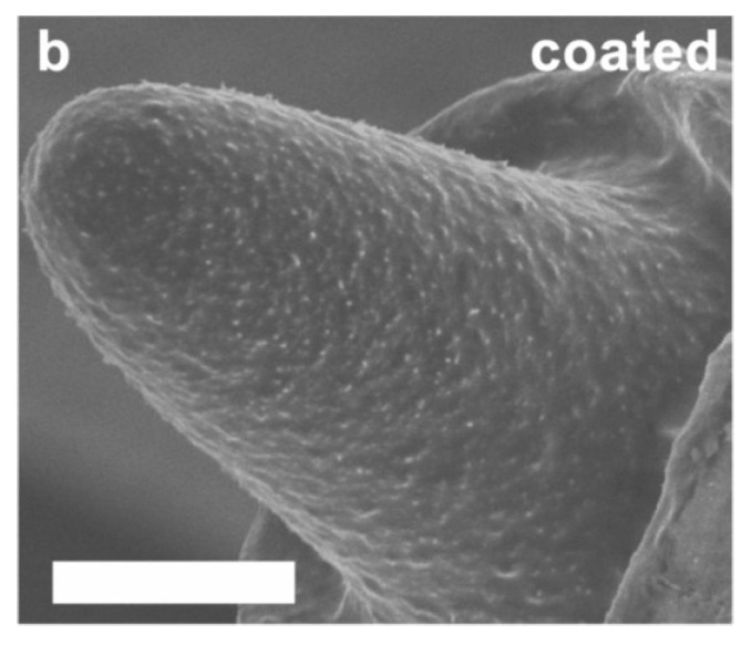
Figure 5.
SEM images of representative uncoated (a) and PEDOT/MWCNT/Dex coated (b) electrode tips extracted from the brain after 11 days. Tips were cleaned using trypsinization and dried before imaging. Note intact coating with no visible cracks or spallation, and the presence of a dense biological film overlaying the coating. Scale bars = 3 µm.


