FIGURE 6.
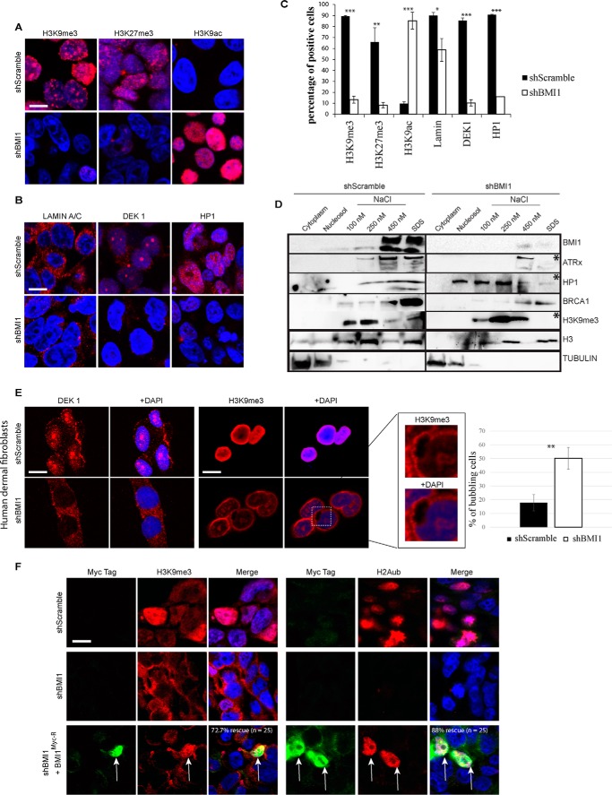
BMI1 knockdown cells present heterochromatin and nuclear envelope alterations. A–C, formaldehyde-fixed 293FT cells were immunolabeled and counterstained with DAPI. Scale bar, 10 μm. Positive cells were counted on 4 different images for a total of 200 cells per condition, and the percentage of positive cells was calculated accordingly. t test with two tails, where *, p ≤ 0.05; **, ≤0.01; ***, ≤0.001. Note that the apparent localization of Lamin A/C in the cytosol is the result of Triton X-100 treatment. D, 293T cells were infected with shScramble or shBMI1 viruses and the compartments of the cell were fractionated. Note the reduction (*) of ATRx, HP1, and H3K9me3 in SDS fractions of shBMI1-treated cells. E, human dermal fibroblasts were infected with shScramble or shBMI1 viruses, immunolabeled, and counterstained with DAPI. Note the reduced DEK1 and H3K9me3 labeling, and H3K9me3 localization at the nuclear periphery, in BMI1-deficient cells. Bubbling of the nuclear envelope was also observed (inset); **, p ≤ 0.01. Scale bar, 10 μm. F, human dermal fibroblasts were infected with shScramble or shBMI1 viruses, and next transfected with a plasmid encoding an RNAi-resistant BMI1 Myc-tagged construct. Note the rescue of H2Aub and H3K9me3 nuclear labeling in Myc-positive cells knockdown for BMI1 (arrows). Scale bar, 10 μm.

