FIGURE 5.
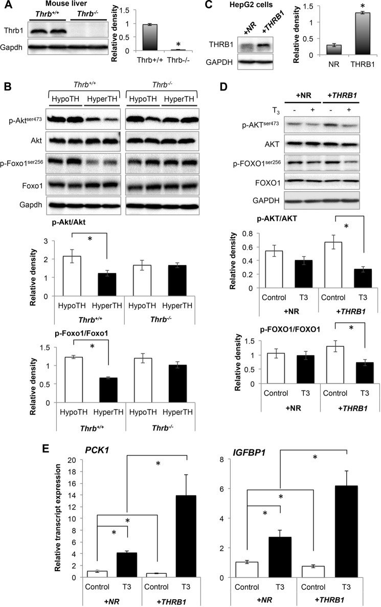
Thyroid hormone (T3) decreased AKT/FOXO1 phosphorylation and increased FOXO1 target gene expression in THRB1-dependent manner. A and B, Western blot analysis to confirm Thrb1 knock-out (A) in mouse liver tissues and phosphorylated Akt/Foxo1 (B) in liver tissues from hypothyroid (HypoTH) and hyperthyroid (HyperTH) of Thrb+/+ and Thrb−/− mice, respectively. Bar graphs below each Western blot data set represent relative densitometric measurements. Statistical significance was calculated as *, p < 0.05, and error bars represent ± S.E. C and D, Western blot analysis to confirm stable overexpression of THRB1 in HepG2 cells (C) and analysis of phosphorylated AKT.FOXO1 in NR-HepG2 and THRB1-HepG2 cells (D). Bar graph represents relative densitometric measurements of phosphorylated AKT and FOXO1. Statistical significance was calculated as *, p < 0.05, and error bars represent mean ± S.D. E, transcript expression of FOXO1 target genes (PCK1 and IGFBP1) by RT-qPCR analysis in NR-HepG2 Versus HepG2 cells expressing THRB1. Statistical significance was calculated as *, p < 0.05, and error bars represent mean ± S.D.

