
FIGURE 6.
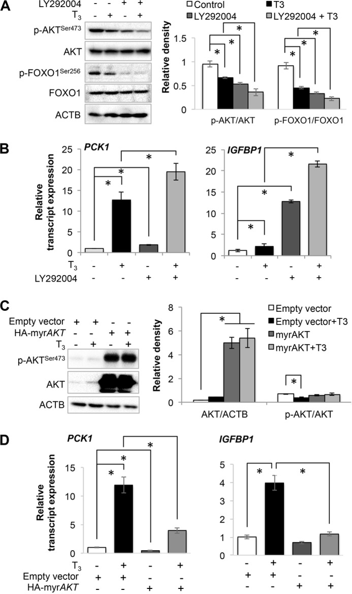
Thyroid hormone (T3) induced FOXO1 target genes in an AKT-dependent manner. A, Western blot analysis for phosphorylated AKT and FOXO1 in HepG2 cells. LY292004 compound (5 μm) was used to inhibit AKT in HepG2 cells along with or without T3 (100 nm). Bar graph represents relative densitometric measurements of phosphorylated AKT and FOXO1. Statistical significance was calculated as *, p < 0.05, and error bars represent mean ± S.D. B, transcript expression of PCK1 and IGFBP1 in HepG2 cells treated with LY292004 compound and/or T3. Statistical significance was calculated as *, p < 0.05, and error bars represent mean ± S.D. C, Western blot analysis of myristoylated AKT (myrAKT), a constitutively active form of AKT, overexpression in THRB1-HepG2 cells. Bar graph represents relative densitometric measurements of total as well as phosphorylated AKT. Statistical significance was calculated as *, p < 0.05, and error bars represent mean ± S.D. D, transcript expression of PCK1 and IGFBP1 in HepG2 cells overexpressing myrAKT and treated with or without T3. Statistical significance was calculated as *, p < 0.05, and error bars represent mean ± S.D. ACTB, β-actin.