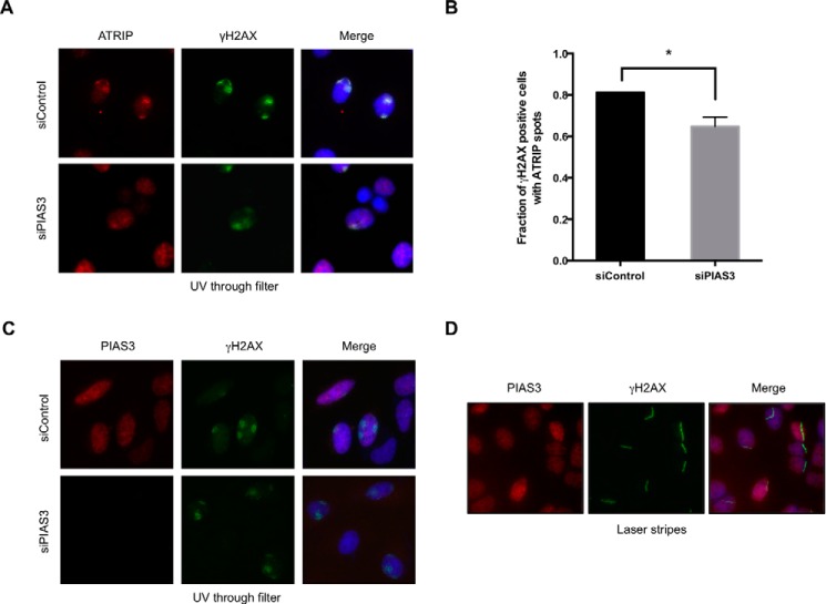
FIGURE 10.
The localization of PIAS3 and its effects on ATRIP recruitment. A and B, HeLa cells transfected with control or PIAS3 siRNA were irradiated with UV (50 J/m2) through filters. The nuclear areas exposed to UV were visualized by γH2AX immunofluorescence. The localization of endogenous ATRIP was analyzed by ATRIP immunofluorescence. Representative images of cells are shown in A. The fractions of irradiated cells (γH2AX-positive cells) with ATRIP spots were quantified (B). *, p < 0.05. C and D, HeLa cells were transfected with control or PIAS3 siRNA. Cells were either irradiated with UV through filters (C) or with UV laser (D). Sites of DNA damage were visualized by γH2AX immunofluorescence. The localization of endogenous PIAS3 was analyzed by PIAS3 immunofluorescence. The staining specificity of the PIAS3 antibody was confirmed by PIAS3 knockdown in C.

