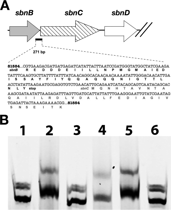
FIGURE 8.
Identification of a DNA-binding site for SbnI. A, schematic identifying the location of a 271-bp DNA fragment that was used for the EMSA experiment illustrated in B. The base numbering is from genome sequence for S. aureus strain Newman. B, electrophoretic mobility shift assay demonstrating SbnI binding to DNA fragment shown in A. Each sample contained 120 ng of fluorescently labeled, 270-bp dsDNA, as well as the following: lane 1, DNA alone; lane 2, 25 μm SbnI; lane 3, 25 μm denatured SbnI; lane 4, 25 μm SbnI with 100:1 unlabeled sbnC probe to labeled probe; lane 5, 25 μm SbnI with 100:1 unlabeled rpoB probe to labeled probe; lane 6, 25 μm SbnG.

