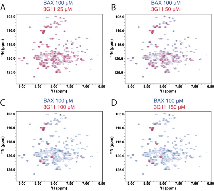
FIGURE 4.
NMR analysis of the 15N-labeled BAX monomer upon 3G11 titration. A–D, NMR HSQC analysis of 15N-labeled BAX (100 μm) upon titration of Fab 3G11 up to a ratio of 1:1.5 BAX:3G11, as indicated for each overlaid spectrum of unbound BAX and 3G11-bound BAX. Unbound BAX HSQC spectra are shown with blue cross-peaks, and 3G11-bound BAX HSQC spectra are shown with red cross-peaks. Dose-dependent loss of the intensity of HSQC cross-peaks of 15N-labeled BAX spectra was observed upon 3G11 binding to BAX. At a 1:1 ratio of BAX:3G11, the vast majority of 15N-labeled BAX cross-peaks disappear because of the formation of a stoichiometric 3G11-BAX complex. No further changes in the 15N-labeled BAX spectra are observed at 1:1.5 ratio of BAX:3G11.

