
FIGURE 8.
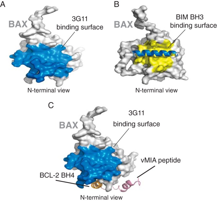
Overlap of the 3G11-binding surface and the BIM BH3-binding surface with the N-terminal trigger site of BAX. A, surface representation of BAX (light gray) highlighting the 3G11-binding surface to BAX (blue) as determined in this study. B, surface representation of BAX (light gray) highlighting the binding surface (yellow) of BIM BH3 (blue helix) in contact with BAX, as determined previously (PDB code 2K7W). C, surface representation of BAX (light gray) highlighting the 3G11-binding surface to BAX (blue) compared with the binding sites of peptides from cytomegalovirus protein vMIA (pink) and BCL-2 BH4 domain (orange).