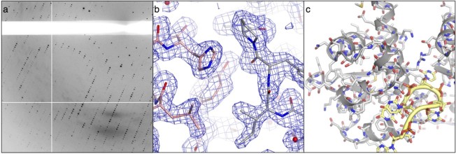
Figure 1 .
Protein crystallography: (a) detail of the X-ray diffraction pattern from a protein crystal, (b) an electron density map, with accompanying atomic model of a protein molecule. Bonds between atoms are shown as sticks, (c) the crystal structure of a protein-RNA complex. Protein and RNA chains are represented by ribbons and tubes respectively. Amino acids and nucleotides are also shown in stick representation (Colour images are available in the online version of this article).

