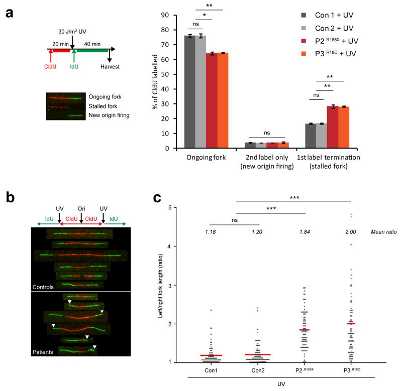
Figure 6. Replication fork stalling is increased following UV-induced DNA damage in TRAIP patient cells.
(a) First label termination events (representing elevated replication fork stalling) are increased after UV irradiation in TRAIP patient fibroblasts. Top left panel, schematic of the experimental plan. Bottom left panel, representative images of ongoing or stalled replication forks and new origin firing from DNA fiber spreads of primary fibroblasts. Right panel, quantification of ongoing forks, 2nd label only (new origin firing) and 1st label termination (fork stalling) structures in fibroblast cells. Mean ± SD, n=3 independent experiments, >400 structures per cell line per experiment quantified. Student’s t-test: *p<0.05; **p<0.01; ns, not significant. (b, c) Substantial fork asymmetry is seen in UV-treated patient cells. (b) Representative images of DNA fibers from controls (Con1, Con2) and patient fibroblasts (P2, P3) after 30 J/m2 UV treatment. (c) Quantification of replication fork asymmetry. Ratio of left/right fork length; mean ratio for each cell line is indicated in italics; Mann Whitney Rank sum test: ***p<0.001; ns, not significant. Data points pooled from n=2 independent experiments, >50 structures per cell line per experiment quantified.

