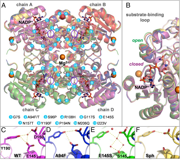
Fig. 5.
(A) Overlay of the crystal structures obtained for apo WT (red), NADP+-bound WT (magenta), A94F (blue), E145S (green), and Sph (yellow). The cofactor is shown as pink sticks; the Mg2+ ions, as orange spheres. Mutations introduced in the studied variants are shown as cyan circles; the active sites, as purple ellipses. (B) The substrate/cofactor binding region showing the open and closed conformations of the loop. (C–F) The Tyr190-Glu/SerS145-Asp150 triad in different variants, showing the water and/or hydrogen bond network.

