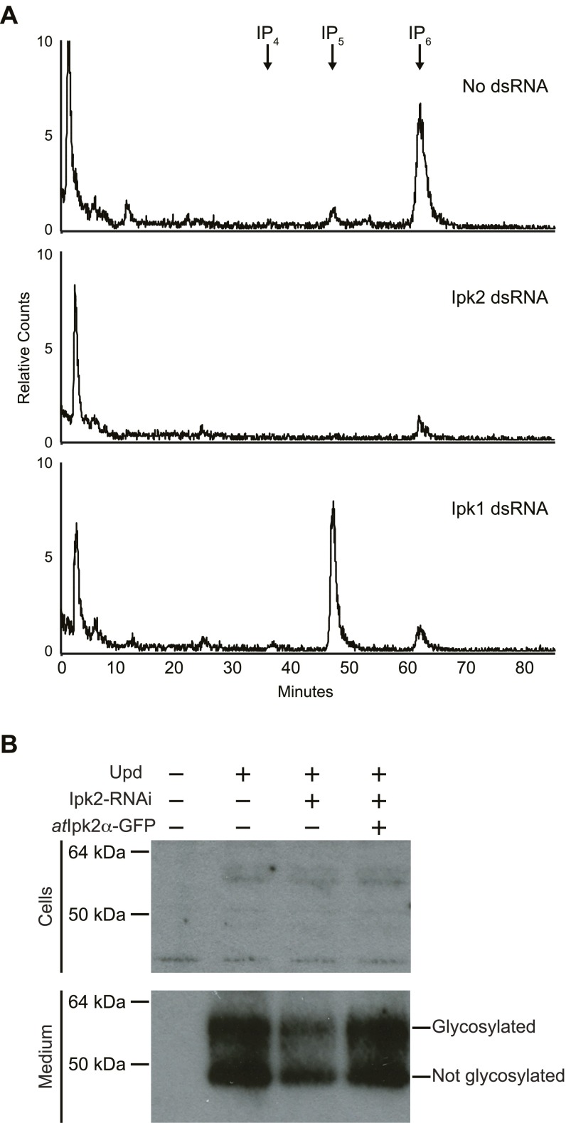
Fig. S4.
(A) Assessment IP levels in Kc167 cells that were treated with dsRNA for Ipk2 or Ipk1. Cells were labeled with [3H]inositol after being treated with dsRNA. Traces show no dsRNA treated control (Top), Ipk2 dsRNA (Middle), or Ipk1 dsRNA (Bottom). Cells were grown in media containing 50 μCi/mL [3H]inositol. Soluble extracts were separated by Partisphere strong-anion exchange HPLC. (B) Ipk2 knockdown causes a reduction in secreted Upd. Cells were transfected with combinations of Upd plasmid, atIpk2-GFP plasmid, and treated with Ipk2 dsRNA as indicated by the + and – signs. Cells were treated with heparin to release Upd from the extracellular matrix. Protein extracts from the treated cells and the conditioned medium were immunoblotted with antibodies against Upd. The top gel shows the cells and the bottom gel shows the conditioned medium. We were unable to detect appreciable amounts of Upd in the cell samples (Top), whereas the conditioned medium appeared to contain the bulk of the Upd (Bottom). The two bands of Upd correspond to the glycosylated and unglycosylated forms. The glycosylated form is shown in Fig. 4B.

