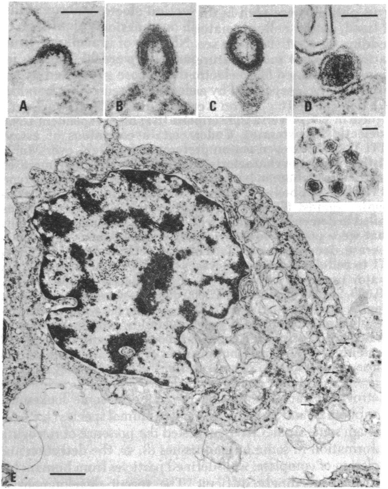
Fig. 1.
Thin section electron micrographs of HUT-102 cells. (A–D) “C-type” particles in various stages of budding and maturation. (E) IUdR-induced cell showing large numbers of budding and released particles, some of which are enlarged in the Inset. (Scale bars, A–D and Inset, 100 nm; E, 1,000 nm.) Reproduced with permission from ref. 19.

