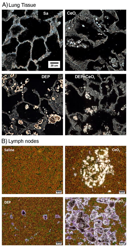
Fig. 7.
CeO2 particles in the lung tissue and lymph nodes collected from particle-exposed rats, at 28 days post-exposure. Control lung tissues (A) and lymph nodes (B) exhibit no particles under high resolution, dark field illumination. Illuminated CeO2 particles, using enhanced darkfield-based illumination, were clearly detected in the macrophages, in the interstitium, in the acellular surfactant clumps, and in the airspace of the CeO2 (7 mg/kg)- and DEP (35 mg/kg) + CeO2-exposed lungs at 28 days after exposure. As shown in the representative micrographs, lymph nodes of CeO2-exposed lungs contained isolated clusters of particle accumulations. Lymph nodes of DEP-exposed lungs demonstrated isolated, dense clumps of particles, while the combined exposure (CeO2 + DEP) resulted in nearly continuous particle accumulations throughout the lymph node.

