Figure 1.

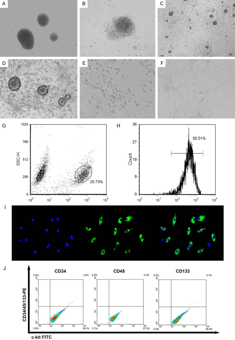
Isolation and flow cytometric sorting of c-kit+ CSCs. A. There was virtually no detectable outgrowth 24 h after plating. B. CEM stimulated the extensive outgrowth of cells, and only a small number of isolated cells were observed in the culture dish at 72 h. C and D. Most 7 day rat heart explants exhibited cellular outgrowth from the edge of the explants. E. FACS-isolated c-kit+ CSC cells from cardiac explants were replanted in CGM. F. Isolated cells were plated onto glass coverslips, which had been previously coated with type I collagen from rat tail. Angiotensin II could induce c-kit+ CSC cells into cardiomyocyte-like cells. G. Cells were isolated from cardiac explants and subjected to FACS as described in Methods section. Gating of c-kit+ CSCs. H. Representative post-sorting histogram identifying c-kit+ CSCs. After culture for 48 h, the c-kit-labeled cell population routinely constituted ~95.51% of the total gated cells. I. Immunocytochemistry of stained c-kit+ CSCs. These passage cells were incubated overnight with primary c-kit monoclonal antibodies (1:1500 dilution). The cell layers were then rinsed with PBS and incubated with FITC-conjugated anti-rabbit antibodies (1:200 dilution). Nuclei were counterstained with DAPI. J. These cells were positive for the expression of cell-surface marker c-kit and negative for expression of each one of cell-surface markers CD34, CD45, and CD133.
