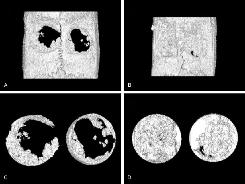
Figure 4.

Reconstructions of bone defects using micro-CT. Bone defects were covered by SF nanofibrous membranes (left) and Bio-Gide® membranes (right). (A) The front image of operated specimen at 4 weeks; (B) The front image of operated specimen at 12 weeks; (C) The ROI image of operated specimen at 4 weeks. (D) The ROI image of operated specimen at 12 weeks. CT, computed tomography; ROI, region of interest; SF, silk fibroin.
