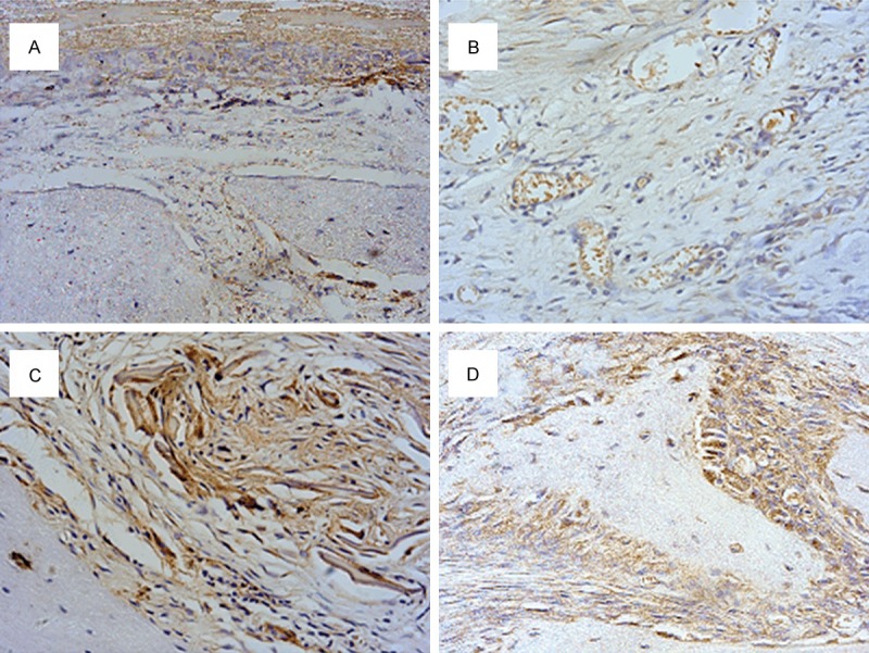
Figure 7.

Collagen I antibody immunohistological staining of rat calvarial defects covered by SF nanofibrous membranes or Bio-Gide® membranes after 4 and 12 weeks. (A) SF nanofibrous membrane at 4 weeks postoperatively; (B) Bio-Gide membranes at 4 weeks postoperatively; (C) SF nanofibrous membranes at 12 weeks postoperatively; (D) Bio-Gide membranes at 12 weeks postoperatively (magnification, × 400). SF, silk fibroin.
