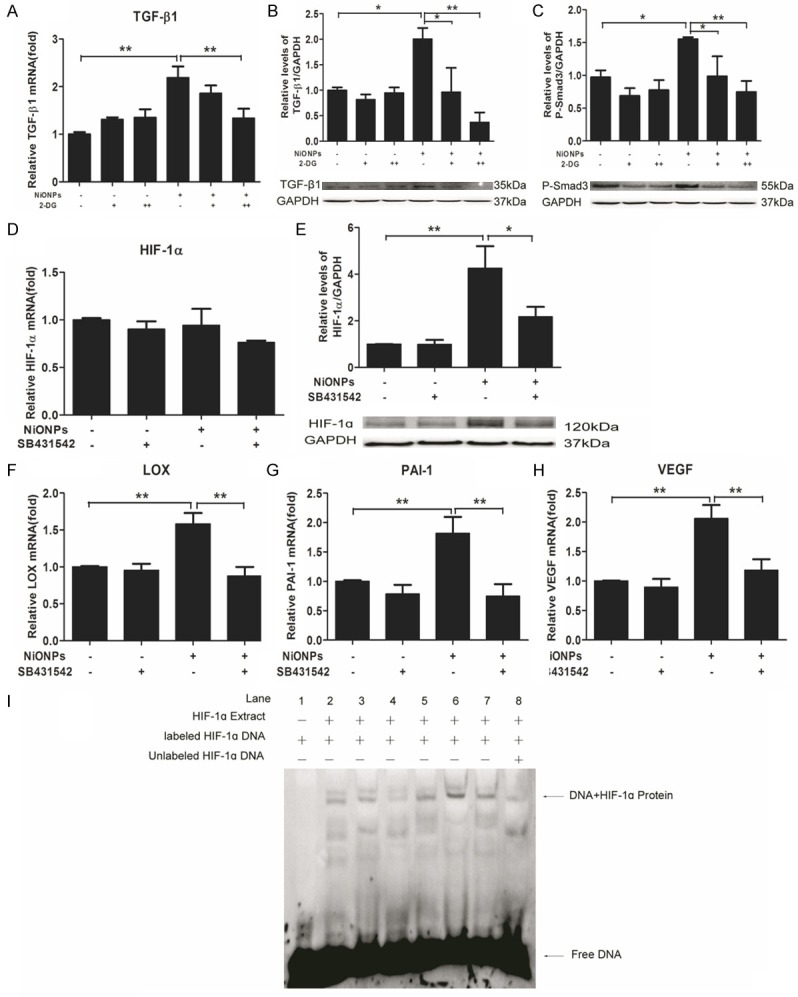
Figure 5.

Reciprocal regulation between HIF-1α and TGF-ß1. (A-C) Human fetal lung fibroblasts were pretreated with 10 mM (+) and 20 mM (++) 2-DG (HIF-1α inhibitor) for 2 h and then exposed to 2 μg/cm2 NiONPs for 24 h. (D-H) Human fetal lung fibroblasts were treated with 10 nM SB431542 and 2 μg/cm2 NiONPs simultaneously for 24 h. The levels of TGF-ß1 (A), HIF1α (D), LOX (F), PAI-1 (G) and VEGF (H) were detected by qPCR and Protein expressions of TGF-ß1 (B) and P-Smad3 (C) were analyzed by western blot (B) and normalized to GAPDH. (I) EMSA was performed to analyze the binding of proteins from nuclear extracts of human fetal lung fibroblasts treated with 2 μg/cm2 NiONPs plus 10 nM SB431542 or 1 ng/ml TGF-ß1 for 24 h to the consensus HIF-1α binding site. Lane: 1, negative control; 2, ctl; 3, TGF-ß1; 4, SB431542; 5, NiONPs; 6, NiONPs+TGF-ß1; 7, NiONPs+SB431542; 8, cold-competitive control.
