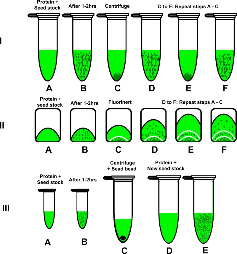
FIG. 4.
A schematic representation of three different seeding protocols. I: Multiple seeding: A. Mixing the seed stock with protein. B. After 1–2 h. C. Centrifuging the crystal suspension at 3000 rpm for 10 min and collecting the supernatant to a new tube. D. After adding the 1:10 seed stock (that has 10% PEG 2000 in buffer A with 0.013% βDM). E and F are repeating C and D to get another wave of dPSIIcc microcrystals. II: In situ multiple seeding: A. The crystallization setup. B. After 1–2 h. C. After addition of Fluorinert to separate the microcrystals from the next wave. D. After addition of seeding stock and increasing the concentration of PEG 2000. E and F. Repeating the same procedure as C and D for the subsequent dPSIIcc microcrystals waves. III: Double seeding: A and B are the same as A and B for multiple seeding. C. The microcrystals from B were collected and crushed using seed bead kit from Hampton research. D. The crushed crystals were used to prepare a 500 μl seed stock. D. 408 μl of this seed stock was mixed with 96 μl of dPSIIcc protein solution of concentration 40 mg/ml (4 mM Chla).

