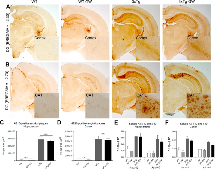
Fig 2. GW3965 treatment did not produce detectable reduction in amyloid beta load.
Amyloid plaque deposition in brains of treated and untreated mice groups were evaluated by immunohistochemistry and soluble Aβ (1–42) by ELISA. (A-B) Representative micrographs of brain sections of Aβ immunohistochemistry with antibody anti-Aβ (6E10). (C-D) No significant differences were observed on the anti-Aβ positive area in the hippocampus or cortex of treated 3xTg-AD mice. Data were expressed as mean ± S.E.M. Statistical analysis was done by one way ANOVA followed by Tukey's multiple comparison test. Females n = 4 per 3xTg-AD group. (E-F) RIPA soluble Aβ42 and Aβ40 in the hippocampus or cortex were measured by ELISA. Quantification data were expressed as mean ± S.E.M. Statistical analysis was done by one way ANOVA followed by Tukey's multiple comparison test. n = 5 per group.

