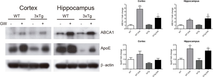
Fig 4. Brain ApoE and ABCA1 protein levels were up regulated by LXR agonist treatment.
Brain levels of LXR target genes were evaluated in the hippocampus and cortex by western blot analysis. (A) Representative images of ApoE, ABCA1 and β-actin Western Blot detection in cortex and hippocampus in WT or 3xTg-AD mice treated with either vehicle or GW3965. (B) Densitometric quantification of Western blot assay of ApoE and ABCA1 in brain lysates of cortex and hippocampus from WT or 3xTg-AD mice treated with either vehicle or GW3965. Samples from each mouse were normalized to actin and expressed as fold-change. Data is expressed as mean ± S.E.M. Statistical analysis was done by one way ANOVA with Tukey's multiple comparison test, n = 5 per group. ***: p < 0.001; *: p < 0.05 compared with WT; #: p < 0.05; ##: p < 0.01 compared with untreated 3Tg-AD.

